We will be presenting our two papers on Head and Neck tumor segmentation and prognostic prediction, and one paper on colorectal cancer detection in whole slide images at MICCAI workshops Sept. 27th and Oct. 1st. Together with our paper at the main conference and the organization of the HECKTOR challenge, we are happy to have a total of five great contributions to MICCAI this year.
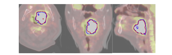
- Our paper on Multi-Task Deep Segmentation and Radiomics for Automatic Prognosis in Head and Neck Cancer will be presented by Vincent Andrearczyk at PRIME 2021.
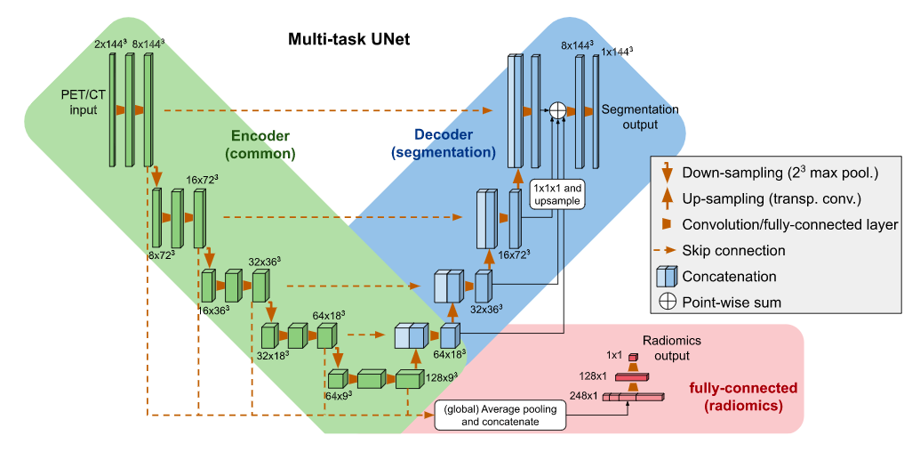
- Our other paper on Fully Automatic Head and Neck Cancer Prognosis Prediction in PET/CT will be presented by Pierre Fontaine at ML-CDS 2021.
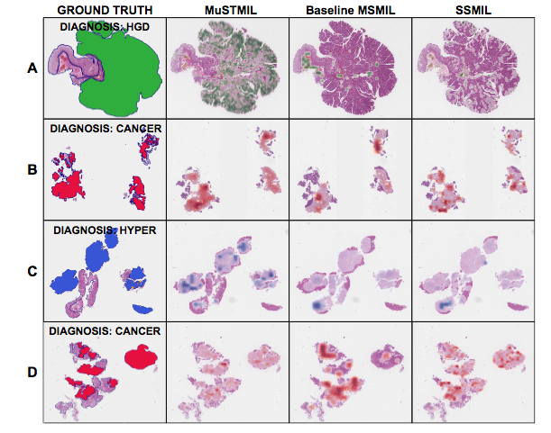
- Our third workshop paper on Multi-Scale Task Multiple Instance Learning for the Classification of Digital Pathology Images with Global Annotations will be presented by Niccolò Marini at COMPAY 2021