In addition to last year’s challenge, we will include more data for the segmentation task as well as a new task for the automatic prediction of patient outcome.
Evaluate your cutting edge algorithm on 300 cases with high-quality annotations from six centers.
Our work “On the Scale Invariance in State of the Art CNNs Trained on ImageNet”, by Mara Graziani, Thomas Lompech, Henning Müller, Adrien Depeursinge and Vincent Andrearczyk. was published with open access rights in a special issue of the Machine Learning and Knowledge Extraction (MAKE) journal: https://www.mdpi.com/2504-4990/3/2/19#abstractc
By using our tool Regression Concept Vectors (pip install rcvtool), we modeled information about scale within intermediate CNN layers. Scale covariance peaks at deep layers and invariance is learned only in the final layers. Based on this, we designed a pruning strategy that preserves scale-covariant features. This gives better transfer learning results for medical tasks where scale is discriminative, for example, to distinguish the magnification level of microscope images of tissue biopses.
The second edition of the HECKTOR challenge was accepted at MICCAI 2021. This challenge is designed for researchers in medical imaging to compare their algorithms for automatic segmentation of tumor in Head and Neck Cancer. In addition to last year’s challenge, we will include more data for the segmentation task as well as a new task for the automatic prediction of patient outcome.
This week took place the kickoff meeting of BIGPICTURE, an EU project gathering about 50 partners including hospitals, research centers and pharmaceutical companies with the aim to create the largest database of histopathology images for computer assisted cancer diagnostic and treatment planning.
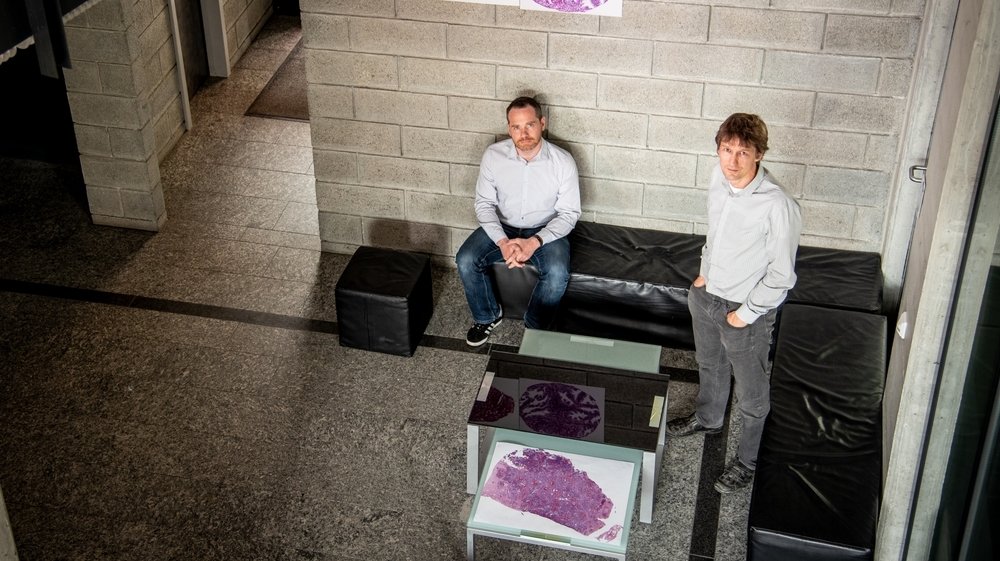
Manfredo Atzori, senior researcher and Henning Müller, professor at the Institute of Information Systems, HES-SO
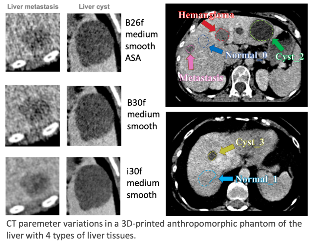
Our abstract entitled ‘Assessment of the stability and discriminative power of radiomics features in liver lesions using an anthropomorphic 3D-printed CT phantom’ by Jimenez-del-Toro et al. has been accepted to be presented at the Swiss Congress of Radiology 2021 ONLINE edition, as an Oral Presentation.
Our abstract entitled ‘Revealing the most suitable CT radiomics features for retrospective studies with heterogeneous datasets’ by Jimenez-del-Toro et al. has been accepted to be presented at the ONLINE European Congress of Radiology 2021, for the session ‘AI in abdominal imaging’, held from March 3-7, 2021.
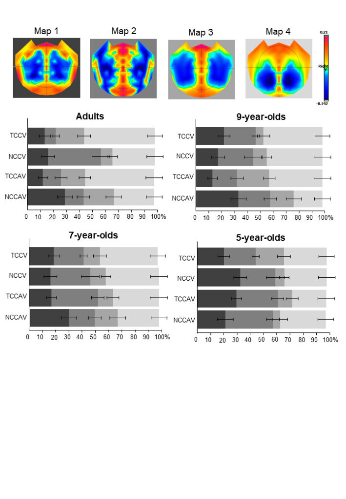
This work led by Nora Turoman, Pawel J. Matusz and their colleagues focused on using advanced multivariate EEG analyses to understand how children develop their attentional control in real-world like settings and when different processes that underlie this attention control reach the adult-like state.
Our team, led by Prof. Henning Müller, started a collaboration with the American College of Radiology to develop AI tools that help radiologists in their medical decision making. Le Nouvelliste wrote an article about this collaboration (in French).
We hosted Iam Palatnik (Pontifical Catholic University of Rio de Janeiro, Brazil) at MedGIFT for an invited talk on interpretability in medical imaging. Iam shared challenges and solutions for the Local Interpretable Model Agnostic Explanations (LIME) method. The video of his presentation is available online.
Abstract: An application of Local Interpretable Model Agnostic Explanations (LIME) is described for two case studies: Metastases and Malaria classification. Some of the key challenges of using LIME for this purpose – most notably the instability of explanations – are discussed, as well as some potential solutions. Namely, a genetic algorithm based solution called EvEx, where explanations are evolved as the average of a Pareto Front, and Squaregrid, a parameterless rough approximation. The results seem to show that EvEx finds more consistent explanations for regions of high explanatory weight, and that Squaregrid could be a viable way to diminish the need for segmentation parameter fine tuning.
Our paper on “Breast Histopathology with High-Performance Computing and Deep Learning” (M. Graziani et. al) has been accepted for publication in Computing and Informatics, in the special issue on Providing Computing Solutions for Exascale Challenges.
In this work, we present our modular pipeline for detecting tumorous regions in digital specimens of breast lymph nodes with deep learning models. We evaluate challenges and benefits of training models on high-performance and cloud computing with millions of images.
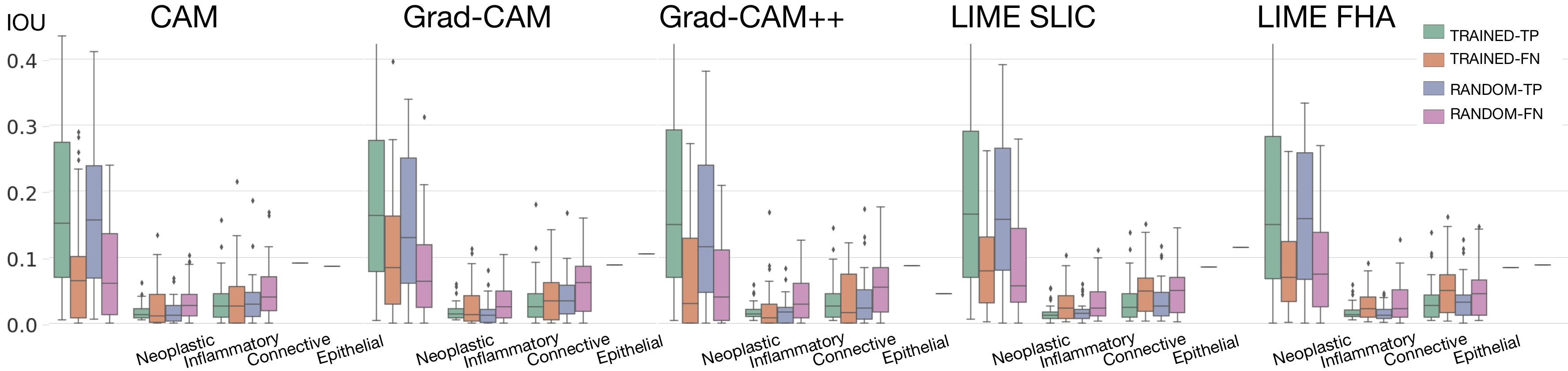
Our PhD student Mara Graziani discussed the topic of defining machine learning interpretability at CIBM (Center for Biomedical Imaging, Switzerland). She also presented our latest applications to the histopathology domain. In particular, she covered our recent work on the “Evaluation and Comparison of CNN Visual Explanations for Histopathology”. She then explained how interpretability can be used in a proactive way to improve model performance. You can watch her talk online at this link: https://www.youtube.com/watch?v=7hs21U-3hgk&feature=youtu.be
Below, the information about the presentation:
Title: A myth-busting attempt for DL interpretability: discussing taxonomies, methodologies and applications to medical imaging.
Abstract:
Deep Learning (DL) models report almost perfect accuracy on some medical tasks, though this seems to plunge in real-world practices [1]. Started in 2009 as the generation of deep visualizations [2, 3], the field of interpretability has grown and developed over the years, with the intent of understanding why such failures happen and discovering hidden and erroneous behaviors. Several interpretability techniques were then developed to address the fundamentally incomplete problem of evaluating DL models on the sole task performance [4].
While defining the key terms used in the field, I will try to bust some myths on DL interpretability: are explainable and interpretable the same thing? Is a “transparent” model an “interpretable” model? Besides, within the application in the field of medical imaging, I will describe the risk of confirmation bias and present our work on evaluating the reliability of interpretability methods. Finally, I will bring examples from our previous works on how interpretable AI can be used to improve model performance and reduce the distance between the clinical and the DL ontologies.
[1] Yune, S., et al. “Real-world performance of deep-learning-based automated detection system for intracranial hemorrhage.” 2018 SIIM Conference on Machine Intelligence in Medical Imaging: San Francisco (2018).
[2] Erhan, D., et al. “Visualizing Higher-Layer Features of a Deep Network.” (2009).
[3] Zeiler, Matthew D., and Rob Fergus. “Visualizing and understanding convolutional networks.” European conference on computer vision. Springer, Cham, 2014.
[4] Doshi-Velez, Finale, and Been Kim. “Towards A Rigorous Science of Interpretable Machine Learning.” stat 1050 (2017): 2.
Two papers have been accepted for presentation at the 43rd European Conference on Information Retrieval (ECIR 2021), that will take place online online March 28 – April 1, 2021.
The “Germaine de Staël” program promotes collaboration between French and Swiss researchers and research teams. Several exchanges between our group (Prof. Henning Müller) and Sorbonne university of histopathology image analysis (Nicolas Lomenie and Camille Kurtz) will be funded by this project to work on deep learning in digital pathology with gigapixel images of hepatic tissue: “BioGigaDeep -Apprentissage profond en pathologie digitale pour l’analyse d’images gigapixels de tissus hépatiques”.
Our paper entitled “InvNet: A Deep Learning Approach to Invert Complex Deformation Fields”, by Marek Wodzinski and Henning Müller, has been accepted for presentation at ISBI 2021, the IEEE International Symposium on Biomedical Imaging to be held virtually on April 13-16, 2021.
Visualization of the results of the proposed deep learning-based algorithm for inverting complex, nonrigid deformation fields based on a modified U-Net architecture and the symmetric inverse consistency
Prof. Henning Müller will give a keynote presentation on “Multimodal Medical Data Analysis: Machine Learning in Histopathology” at the MMDLCA workshop at the International Conference on Pattern Recognition (ICPR) 2021, on January 11, 12:00 CET.
Congratulations to Marek Wodzinski and Henning Müller for their 2nd Best Paper Award at the International Workshop on Biomedical Image Registration WBIR 2020 with their paper on “Learning-based affine registration of histological images“.
In this work, we evaluate the alignment of XAI visualisations to cancer diagnostic procedures for breast tissue. Do the visualizations highlight specific nuclei types? Visual explanations may induce confirmation bias about CNN decisions.
Quantification of CNN attention on the nuclei types (+ link to the GitHub repository)