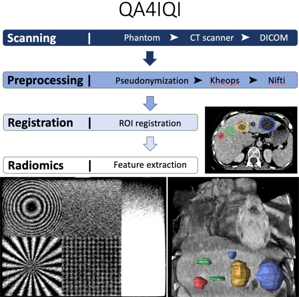
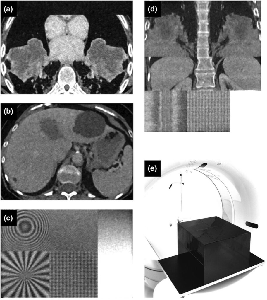
Congratulations to Nataliia Molchanova who received the Summa Cum Laude Merit Award at the recent 2023 ISMRM & ISMRT Annual Meeting & Exhibition for the abstract “Towards Informative Uncertainty Measures for MRI Segmentation in Clinical Practice: Application to Multiple Sclerosis”.
This recognition indicates an emerging interest in Safe AI supporting medical imaging analysis, highlighting an effort of the organizers to raise awareness about the limitations of deep learning and the necessity of moving towards more trustworthy, explainable, and transparent AI solutions in clinical practice.
Nataliia Molchanova and her work on uncertainty measures for MRI segmentation
Niccolò Marini successfully defended his PhD work on “Deep learning methods to reduce the need for annotations for the extraction of knowledge from multimodal heterogeneous medical data”. His thesis focused on:
how to alleviate the need for manual annotations to train deep learning algorithms
how to leverage color variability to improve the CNN generalization on unseen data
how to learn a more details WSI representation combining multiple magnification levels
how to empower the raw-pixel level image representation introducing high-level concepts from textual reports
This work was co-supervised by Prof. Henning Müller and Prof. Manfredo Atzori, Institute of Informatics, University of Applied Sciences Western Switzerland (HESSO), Prof. Stephane Marchand-Maillet, Department of Computer Science, University of Geneva. Prof. Anne Martel, Department of Medical Biophysics, University of Toronto, completed the committee as international expert in the domain.
Cristina Simon-Martinez was interviewed by Research Works to present the ReHaBot project in a very interesting podcast.
RehaBot is a chatbot between therapists and patients to establish tele-rehabilitation programs and quantify their outcome. This project, led in collaboration with Mario Martinez-Zarzuela, received a warm welcome by research and clinical partners.
Learn more about the project in the podcast available online.
The project “FishLab – Semi-autonomous system for centralised video-monitoring of fish migration in river systems” has been granted an InnoCheque funding by Innosuisse. This will support our collaboration with FishLab to develop a standardized/automated observatory of fish flows in rivers.
For this purpose, fish ladders are fitted with underwater cameras with infrared backlighting.
The end customers are dam operators, who are required to demonstrate the ecological restoration of their structures by 2030 (Federal Water Protection Act 2011).
At HES-SO, we are studying video processing for the detection of fish, their tracking, count, species identification and size measurement. We are also developing an interface to check and complete the automated results.
Our article “A dexterous hand prosthesis based on additive manufacturing”, M. Atzori et al., was accepted at the VIII congress of the Italian Group of Bioengineering, which will be held in June 2023.
The article presents a project that has been ongoing for several years now and that is aimed at the development of low cost and dexterous prosthetic hands to be used in real life conditions. The tests of the prosthetic hand demonstrate its dexterity and the potential of future systems based on 3D printing and machine learning. More details are available in the paper.
3D printed robotic hand and socket.Examples of hand grasp tests.
Breaking down a deep learning model into interpretable units allows us to better understand how models store representations. However, the occurrence of polysemantic neurons, or neurons that respond to multiple unrelated features, makes interpreting individual neurons challenging. This has led to the search for meaningful directions, known as concept vectors, in activation space instead of looking at individual neurons.
In this work, we propose an interpretability method to disentangle polysemantic neurons into concept vectors consisting of linear combinations of neurons that encapsulate distinct features. We found that monosemantic regions exist in activation space, and features are not axis aligned. Our results suggest that exploring directions, instead of neurons may lead us toward finding coherent fundamental units. We hope this work helps bridge the gap between understanding the fundamental units of models, an important goal of mechanistic interpretability, and concept discovery.
The proposed method to obtain two concept vectors from a neuron is depicted for a neuron that activates for both apples and sports fields. See full paper for details.
QuantImage v2 (QI2) is an open-source web-based platform for no-code clinical radiomics research developed in collaboration with HES-SO and CHUV. It was developed with the aim to empower physicians to play a leading role in clinical radiomics research.
Physician-centered radiomics research. Physician-centered radiomics envisions medical doctors at the center of the radiomics research, development, and translation cycle
The web page describing QuantImage is available here.
To test and validate novel CT techniques, such as texture analysis in radiomics, repeat measurements are required. In this work, we analyze in detail the quantitative characteristics of iodine-ink based 3D-printed phantom. Design considerations and the manufacturing process are described. Potential pitfalls for radiomics testing for such phantoms are presented.
CT datasets of the different phantom parts and overall phantom appearance. (a) Transversal CT image of the lung-part, (b) the liver part and (c) the test-patterns. (d) coronal CT image of the phantom. (e) cuboid black phantom positioned on a board in a CT scanner.
The Springer LNCS proceedings of the Third Challenge, HEad and neCK TumOR segmentation and outcome prediction in PET/CT images (HECKTOR) 2022, Held in Conjunction with MICCAI 2022, are now published and available online.
The editors V. Andrearczyk, V. Oreiller, M. Hatt, and A. Depeursinge gathered:
23 papers from participants reporting their methods on the tasks of tumor segmentation and outcome prediciton.
An overview paper presenting the data, participation, results etc.
Post-challenge participation is still open on grand-challenge.
The first AI-Cafe of 2023 will put in the spotlight the ExaMode project on February 8th 2023 14-15h CEST.
Prof. Henning Müller will present “ExaMode: a practical approach towards using machine learning in histopathology”. The ExaMode project is funded by the European Commission to work on scalable solutions for AI in histopathology. The project thus follows a practical approach defining use cases, collecting multimodal data from hospitals and and then developing decision support tools. Aspects such as learning from limited data and multimodal learning will be addressed as well as working on aspects of explainability when building systems that are integrated into the clinical workflow.
In this work, we present a method to improve the efficiency of color augmentation methods by increasing the reliability of the augmented samples used for training computational pathology models. During CNN training, a database including over 2 million H&E color variations collected from private and public datasets is used as a reference to discard augmented data with color distributions that do not correspond to realistic data.
This Collection presents a series of articles describing annotated datasets of medical images and video. Data are presented without hypotheses or significant analyses to support improvements such as benchmarking or improving machine learning algorithms. All medical specialities are considered and data can be derived from study participants, tissue samples, electronic health records (EHRs) or other sources. All described datasets are assessed to ensure their open availability (where possible) or secure access controls (where required) via Scientific Data‘s editorial and peer review processes.
Our work on “Mapping of the Upper Limb Work-Space: Benchmarking Four Wrist Smoothness Metrics“, led by Alessandro Scano, Cristina Brambilla, Henning Müller and Manfredo Atzori has been published in the Applied Sciences journal, MDPI.
Smoothness-based models describe effectively skilled motion planning. Despite smoothness being often used as a measure of motor control and to evaluate clinical pathologies, so far, a smoothness map is not available for the whole workspace of the upper limb. In this work, we provide a map of the upper limb workspace comparing four smoothness metrics: the normalized jerk, the speed metric, the spectral arc length, and the number of speed peaks.
Scheme of the work. Upper limb movements (P2P and EXP) were performed in 5 sectors of the workspace by 15 subjects. The data were segmented in phases and the wrist kinematics and the articular angels were computed. Then, four smoothness metrics based on wrist motion were computed and smoothness was compared between sectors, mapped with respect to articular angles and in the 3D workspace for each of the smoothness metric. Finally, the correlations between pairs of metrics were computed.
A 4-pages article has been published in RSVIPVision, highlighting our work on A global taxonomy of interpretable AI, based on an excellent interview of Mara Graziani and Vincent Andrearczyk by Ralph Anzarouth.
More details on our work “A global taxonomy of interpretable AI: unifying the terminology for the technical and social sciences” are available in the paper.
This work was made possible thanks to the large collaboration of experts in various domains: Mara Graziani, Lidia Dutkiewicz, Davide Calvaresi, José Pereira Amorim, Katerina Yordanova, Mor Vered, Rahul Nair, Pedro Henriques Abreu, Tobias Blanke, Valeria Pulignano, John O. Prior, Lode Lauwaert, Wessel Reijers, Adrien Depeursinge, Vincent Andrearczyk and Henning Müller.
Pierre Fontaine successfully defended his PhD work on “Extraction and analysis of image features within a radiomics work-flow applied to prediction in radiotherapy”. His thesis focused on how to optimally exploit regions of interest in medical images when building radiomics models for prognosis prediction. His list of publications is available online: http://publications.hevs.ch/index.php/authors/show/2474
This work was co-supervised by
Dr Oscar Acosta, and Prof. Renaud De Crevoisier, Univ Rennes, CLCC Eugene Marquis, INSERM, LTSI – UMR 1099, F-35000 Rennes, France
Prof. Adrien Depeursinge, Institute of Informatics, University of Applied Sciences Western Switzerland (HESSO).
Valentin Oreiller succesfully defended his PhD thesis on Validating and Optimizing the Specificity of Image Biomarkers for Personalized Medicine. In his work, supervised by Prof. Adrien Depeursinge (HES-SO) and, Prof. John O. Prior (CHUV), Valentin obtained excellent achievements:
He designed directional image operators that are Locally Rotation Invariant (LRI), based on the power spectrum and bispectrum of the circular harmonics expansion for 2D images or spherical harmonics expansion for 3D images.
He also tackled a crucial aspect in designing robust predictive models, that is the need for large datasets and benchmarks. An important part of his thesis is the organization of the HEad and neCK tumOR segmentation and outcome prediction in PET/CT images (HECKTOR) challenge.
The quality and reproducibility of radiomics studies are essential requirements for the standardisation of radiomics models. As recent data-driven respiratory gating (DDG) [18F]-FDG has shown superior diagnostic performance in lung cancer, we evaluated the impact of DDG on the reproducibility of radiomics features derived from [18F]-FDG PET/CT in comparison to free-breathing flow (FB) imaging.
This study showed that 131 out of 141 radiomics features calculated on all pulmonary lesions can be used interchangeably between DDG and FB PET/CT acquisitions. Besides, radiomics features derived from pulmonary lesions located inferior to the superior lobes are subject to greater variability as well as pulmonary lesions of smaller size.
Our review article “Automated Tumor Segmentation in Radiotherapy“, by Ricky Savjani et al. has been published in Seminars in Radiation Oncology. In this work, we present advances in gross tumor volume automatic segmentation made in multiple key sites: brain, head and neck, thorax, abdomen, and pelvis.
Automatic tumor segmentation can decrease clinical demand, provide consistency across clinicians and institutions for radiation treatment planning. Additionally, automatic segmentation can enable imaging analyses such as radiomics to construct and deploy large studies with thousands of patients.
(A) In this review, we highlight major advances in tumor autosegmentation for 5 major clinical sites: brain, head and neck, thorax, abdomen, and pelvis. (B) Successful autosegmentation models rely on several steps including: data collection and curation, pre-processing and data ingestion, splitting datasets into train/validate/test sets, hyper-parameter optimization and tuning, architecting networks, post-processing and visualization, and aggregating outputs from ensembles of networks.
The satellite event of HECKTOR (HEad and neCK TumOR segmentation and outcome prediction) at MICCAI 2022 was a great success in terms of participation and quality.
Congratulations to all participants, and particularly to the winners:
Task 1 (segmentation) winner: Andriy Myronenko et al., team NVAUTO
Task 2 (outcome prediction) winner: Louis Rebaud et al., team LITO
Submissions have now re-opened on grand-challenge, we are looking forward to new submissions.
Find more about the HECKTOR challenge in this amazing post.